Understanding Legg-Calvé-Perthes Disease
Legg-Calvé-Perthes disease is a childhood hip condition in which blood supply to the ball-shaped top of the thighbone (femoral head) is temporarily disrupted, causing the bone tissue to weaken and, in some cases, collapse.
It most commonly affects children between ages four and ten, and while the exact cause is not fully understood, early recognition and appropriate management play a crucial role in preserving normal hip shape and function as the child grows.
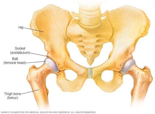
What Happens in the Disease Process?
The femoral head relies on a delicate blood supply, and when this is temporarily interrupted, bone tissue weakens and becomes prone to deformity under normal weight-bearing forces. Over months to a few years, the body gradually restores blood flow and remodels the bone, but if the femoral head becomes misshapen during this process, it can affect long-term hip joint alignment and function.
Signs and Symptoms in Children
- A noticeable limp, which is frequently painless during the early stages
- Hip, groin, thigh, or knee pain that may come and go intermittently
- Reduced hip range of motion, particularly with internal rotation and abduction
- Progressive muscle wasting in the thigh on the affected side over time
- Symptoms that worsen with physical activity and ease with rest
Who Is Most at Risk?
- Boys, who are affected roughly four to five times more frequently than girls
- Children between four and eight years old most commonly, though it can occur outside this age window
- Children who are smaller in stature or exhibit a delayed bone age for their group
- A small proportion of cases presenting with a positive family history
Diagnosis
Diagnosis typically begins with standard X-rays of the hip, which display characteristic structural changes in the femoral head as the disease progresses through its stages.
MRI may be utilized in early cases when X-ray changes are not yet visible, providing a much earlier and more detailed assessment of blood supply impairment and bone involvement.
Treatment Approaches
- Observation: Monitoring and activity modification for younger children with mild involvement
- Physiotherapy: Dedicated physical therapy to maintain hip range of motion and muscle strength throughout treatment
- Bracing: Specialized bracing in select cases to help maintain proper spherical positioning of the femoral head
- Surgical containment: Surgical procedures for older children or more severe cases to preserve joint shape
- Extended monitoring: Long-term clinical follow-up through the healing process, which can take one to several years
Long-Term Outlook
Many children with Legg-Calvé-Perthes disease go on to enjoy good long-term hip function, particularly when diagnosed early and managed appropriately.
Younger age at onset and less severe femoral head involvement are generally associated with a more favorable outcome, though ongoing specialist follow-up remains important throughout bone remodeling.
Frequently Asked Questions
What age does Legg-Calvé-Perthes disease usually affect?
It most commonly appears in children between four and eight years old, though it can occasionally occur in children slightly younger or older than this typical range.
Is Legg-Calvé-Perthes disease caused by an injury?
No specific physical injury is typically identified as the direct cause; it results from a temporary disruption in blood supply to the femoral head, the exact trigger for which is often unclear.
Does every child with this condition need surgery?
No. Many younger children with mild involvement are managed successfully with observation, activity modification, and physiotherapy, with surgery reserved for more severe or older-onset cases.
How long does recovery from Legg-Calvé-Perthes disease take?
The overall bone healing and remodeling process can take one to several years, during which children typically continue with modified activity levels and regular clinical monitoring.
Take Care of Your Child's Hip Health
Concerned about childhood hip pain or looking for a specialist pediatric orthopedic diagnosis? Book a consultation today with our experts.
Book Hip Specialist Consultation