Slip Disc: Pelvic Girdle Pain, Diagnosis and Treatment
A comprehensive guide to understanding pelvic girdle pain links, diagnostic procedures, and targeted treatment options.
Book Free Consultation
Enter your details and our senior medical coordinator will reach out.
Slip Disc and Pelvic Girdle Pain
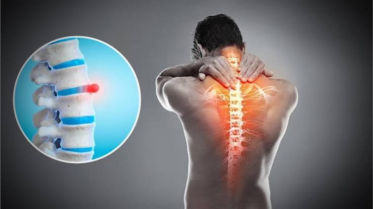
Pelvic girdle pain — discomfort around the pelvis, hips, or lower back — can sometimes trace back to a disc problem at L4-L5 or L5-S1, since nerves from this region also supply the pelvic area.
Pressure on these nerves can make pain radiate into the pelvis or groin, mimicking a purely pelvic issue.
Common Symptoms
Do not ignore these physical indicators associated with slip disc and nerve pressure:
Radiating Pain
Pain radiating from lower back through buttock and down one leg.
Burning Sensation
A sharp, burning, or shooting sensation down the lower extremity.
Nerve Tingling
Tingling or numbness along the sciatic nerve pathway.
Movement Strain
Pain that worsens significantly with sitting, coughing, or sudden twists.
What Causes It?
- Herniated or slipped disc pressing on the nerve root
- Spinal stenosis (narrowing of the spinal canal)
- Degenerative disc disease or bone spurs
- Piriformis syndrome, where a tight hip muscle irritates the nerve
- Spondylolisthesis (a slipped vertebra)
- Pregnancy, obesity, or prolonged sitting, which add pressure on the lower spine
Diagnosing a Slip Disc
Our structured 3-tier comprehensive diagnostic protocol maps over 80 specific clinical history, physical nerve tensioning, and neuroimaging parameters.
1. Medical History & Onset
Detailed recording of nerve radiation, pelvic pain connection, and lifting incidents.
- Radiating Pain Distribution: Mapping pain from lower back through buttock down one leg.
- Pelvic Girdle Link: Tracking nerve symptoms referred into the pelvis and groin from L4-S1 levels.
- Electrical Jolt Onset: Recording sharp, burning, or shooting pain episodes.
- Sitting Tolerance Log: Measuring duration before seated pressure aggravates nerve pain.
- Valsalva Pressure Trigger: Documenting pain spikes during coughing, sneezing, or straining.
- Sensory Loss Reports: Logging patch numbness or pins and needles along nerve pathways.
- Muscle Weakness History: Screening for limb giving-way or foot drop symptoms.
- Lifting Incident Context: Documenting sudden twisting or heavy lifting triggers.
- Unilateral Symptom Check: Verifying if nerve pain affects predominantly one side.
- Previous Disc Flare-Ups: Reviewing historical recurring lower back and leg pain.
- Night Sleep Disruption: Assessing if nerve pain prevents finding a restful sleeping position.
- Cauda Equina Red Flags: Screening for emergency bladder or bowel control issues.
- Previous Treatment Response: Evaluating past epidural injections or physical therapy outcomes.
- Activity Limitation Log: Assessing impact on walking, driving, and working.
- Pain Intensity Scoring: Rating nerve distress levels using standard clinical scales.
- Symptom Progression Rate: Tracking how quickly numbness or weakness has expanded.
- Symptom Triggers: Identifying specific movements worsening disc pressure.
- Associated Fatigue: Screening for general muscle tiredness.
- Weight Stress Factors: Factoring in spinal load.
- Overall Health Overview: Comprehensive medical status summary.
2. Physical Examination
Hands-on nerve stretch testing, reflex evaluation, and muscle power screening.
- Straight Leg Raise (SLR) Test: Provoking compressed nerve roots between 30 and 70 degrees of elevation.
- Slump Test: Assessing neural tension across spinal cord and sciatic pathway in seated posture.
- Deep Tendon Reflexes: Testing knee jerk (L4) and ankle jerk (S1) reflex integrity.
- Myotome Power Testing: Checking muscle strength in foot dorsiflexion and great toe extension.
- Dermatome Sensation Exam: Assessing light touch and pinprick across lower extremity dermatomes.
- Femoral Nerve Stretch Test: Evaluating upper lumbar disc herniation (L2-L4).
- Toe and Heel Walking: Screening for lower limb motor root weakness under weight-bearing.
- Lumbar Range of Motion: Measuring flexion, extension, and side-bending movement restrictions.
- Posture & Antalgic Lean Check: Observing protective spinal shifting away from disc bulge.
- Palpation Screening: Checking paraspinal muscle tenderness and secondary muscle spasm.
- Pelvic Girdle Screening: Evaluating pelvic ring stability to differentiate local joint pain from disc referral.
- Core Stability Test: Evaluating transverse abdominis and back stabilizer endurance.
- Balance Evaluation: Assessing neuromuscular stability during standing tests.
- Transition Observation: Watching patient move between sitting, standing, and lying exam table positions.
- Flexibility Testing: Checking hamstring length.
- Trunk Control Screen: Assessing core support.
- Functional Movement: Observing daily mechanics.
- Stability Check: Assessing segmental control.
- Specialized Tests: Advanced clinical screening.
- Physical Summary: Compiling exam metrics.
3. Imaging & Neurodiagnostics
Lumbar MRI scans, EMG nerve studies, bone structure X-rays, and diagnosis.
- MRI Scan (Gold Standard): Detailed visualization of disc position, annular tears, and nerve root contact.
- Lumbar X-Rays: Assessing bone structure, disc space height, and ruling out fractures.
- CT Scan with Myelogram: High-resolution cross-sectional bone and canal evaluation when MRI is restricted.
- Electromyography (EMG): Testing electrical activity in muscles to confirm chronic nerve root injury.
- Nerve Conduction Study (NCS): Evaluating electrical signal speed across compressed nerve pathways.
- Disc Degeneration Grading: Assessing severity of disc wear and tear on axial and sagittal views.
- Neural Foraminal Stenosis Check: Checking narrowing where nerve roots exit spinal canal.
- Blood Inflammatory Workup: Running ESR and CRP to rule out systemic infection or tumor.
- Differential Diagnosis: Distinguishing discogenic pain from facet joint syndrome, SI joint dysfunction, or piriformis syndrome.
- Diagnostic Selective Nerve Root Block: Confirming precise pain generator via fluoroscopic anesthetic injection.
- Sequential MRI Review: Comparing prior scans to track disc herniation regression over time.
- Surgical Consultation Review: Assessing candidacy for microdiscectomy when conservative care fails.
- Comprehensive Clinical Diagnosis: Synthesizing physical nerve tension tests, history, and MRI findings into a definitive diagnosis.
- Digital Record Archiving: Secure cloud storage of radiological reports.
- Specialist Peer Review: Multi-expert consensus on slip management.
- Risk Stratification: Assessing nerve root compromise risks.
- Prognosis Review: Estimating recovery timelines.
- Lab Correlation: Matching systemic blood markers.
- Bone Density Check: Screening skeletal health.
- Clinical Clearance: Approving targeted therapy start.
4. Treatment & Recovery Plan
Targeted spinal decompression, core rehabilitation, and nerve care.
- Spinal Decompression Therapy: Gentle mechanical traction protocols designed to reduce intradiscal pressure and retract herniated material.
- Targeted Physiotherapy: Specialized core stabilization regimens to protect lumbar discs and improve posture.
- Nerve Gliding Exercises: Controlled stretching movements to ease tension and improve mobility along compressed nerve pathways.
- Pain Management Protocols: Non-invasive modalities and coordinating epidural steroid injections to lower acute nerve root inflammation.
- Ergonomic & Lifting Education: Practical instructions on workstation adjustments, sitting posture, and safe bending mechanics.
- Home Exercise Program: Custom daily stretching routines to maintain spinal flexibility without provoking symptoms.
- Activity Pacing & Modification: Guidance on managing daily tasks and avoiding high-impact bending or twisting.
- Heat & Cold Therapy Guidance: Managing muscle spasms and localized discomfort safely at home.
- Weight Management Support: Reducing mechanical axial load stress on herniated discs.
- Sleep Hygiene Optimization: Pillow placement and mattress support adjustments to relieve nighttime nerve pressure.
- Milestone Progress Tracking: Monitoring recovery and nerve decompression week by week.
- Regular Clinical Follow-ups: Periodic evaluations with senior spine care specialists.
- Patient Self-Care Briefing: Clear instruction on managing slip disc flare-ups independently.
- Long-Term Protection Strategy: Preserving functional independence and active lifestyle freedom.
- Maintenance Care Routines: Sustaining spinal health post-rehabilitation.
- Re-injury Prevention: Safe lifting and bending mechanics training.
- Mobility Enhancement: Safe hip and core movement maintenance.
- Specialist Consult Coordination: Multi-expert review for persistent or severe nerve compression.
- Recovery Milestone Review: Assessing treatment success.
- Active Lifestyle Clearance: Final approval for return to full daily activities.
Treatment Protocols for Conditions
Targeted modalities and structured rehabilitation exercises tailored to specific spinal and pelvic conditions:
1. Slip Disc – L4–L5 / L5–S1
Treatment / Modalities
- Decompression
- TENS: For pain relief
- Manual therapy: Mobilization and targeted muscle release
- Hot pack
- Laser
Rehabilitation Exercises
- McKenzie extension exercises
- Core stabilization
- Pelvic tilts
- Bridging
- Bird-dog
- Neural mobilization
- Hamstring and hip flexibility
- Progressive functional strengthening
2. Disc Sequestration
Treatment / Modalities
- Decompression
- TENS: For pain relief
- Manual therapy: Mobilization and targeted muscle release
- Hot pack
- Laser
Rehabilitation Exercises
- Core stabilization
- Pelvic tilts
- Bridging
- Bird-dog
- Neural mobilization
- Hamstring and hip flexibility
- Progressive functional strengthening
3. Pelvic Girdle Pain
Treatment / Modalities
- TENS
- Laser
- Hot pack
Rehabilitation Exercises
- Pelvic floor activation
- TA / abdominal bracing
- Pelvic tilt
- Gluteal isometric exercises
- Hip abductor strengthening
- Bridging – pain-free range
- Gentle hip mobility
- Functional exercises – sit-to-stand and walking as tolerated
The L4-L5 Spinal Segment
The L4-L5 motion segment represents the anatomical juncture between the fourth and fifth lumbar vertebrae in the lower back. This specific region acts as a primary physical weight-bearing pillar and movement fulcrum, absorbing high mechanical stress during forward flexion, extension, and rotational movements. Because of its intense structural load-bearing role, it is statistically the most frequently affected site for lumbar intervertebral disc herniations and degenerative wear.
- Nerve Root Vulnerability: When a disc herniates or slips at the L4-L5 level, it typically compresses the exiting L5 nerve root.
- Motor Weakness & Foot Drop: Compression here can impair muscle strength required to lift the foot and great toe upward, raising the clinical risk of foot drop or toe-dragging during gait.
- Radiating Symptoms: Triggers persistent dull aches, sharp electrical jolts, or localized numbness radiating from the lower back across the outer shin and into the top of the foot.
The L5-S1 Lumbosacral Junction
The L5-S1 junction serves as the critical lumbosacral transition zone where the lowest movable lumbar vertebra articulates directly with the sacrum. This unique structural boundary experiences maximal shear forces and rotational torque throughout walking, lifting, and posture changes. Structural instability or disc protrusion at this junction directly compromises the foundational stability of the entire lower torso.
- Nerve Root Vulnerability: Protrusions or extrusion events at the L5-S1 level heavily impact and compress the S1 nerve root.
- Reflex & Power Deficits: Can diminish or completely suppress the ankle jerk tendon reflex while lowering calf muscle propulsion power during walking.
- Classic Sciatica Origin: Serves as the primary source of classic sciatic nerve distress, causing deep shooting pain down the posterior thigh, calf, and into the sole or outer edge of the foot.
Disc Sequestration (Free Fragment)
Disc Sequestration is an advanced, severe progression of lumbar disc herniation. In this pathological stage, the inner gelatinous core (nucleus pulposus) completely breaches both the outer annular walls and the posterior longitudinal ligaments, separating entirely from the parent disc to become a free floating fragment within the spinal canal.
Clinical Characteristics & Risks:
- Chemical Nerve Inflammation: The extruded free fragment provokes an aggressive immune and chemical reaction, causing unyielding, severe localized and radiating nerve root pain.
- Radiological Identification: Easily visualized on high-resolution lumbar MRI diagnostics as a detached loose body residing within the vertebral canal separate from the main disc.
- Specialist Medical Tracking: Due to heavy nerve compression risks and potential motor deficits, sequestered discs require rapid medical evaluation and rigorous clinical monitoring.
Pelvic Girdle Pain & Spine Correlation
Pelvic Girdle Pain (PGP) is characterized by deep, aching discomfort experienced around the posterior pelvis, hips, gluteal region, and pubic symphysis. While frequently attributed to local joint laxity, pregnancy, or pelvic trauma, PGP can frequently be triggered, mirrored, or exacerbated by lower lumbar nerve root entrapment from conditions like a slipped disc.
- Shared Neural Pathways: Lower lumbar and sacral nerve roots supply complex sensory and motor feedback loops extending directly into the deep structures of the pelvic girdle.
- Referred Pain Projection: A lower lumbar disc herniation can project sharp or aching pain straight into the hip joint and groin area, successfully masquerading as primary hip or pelvic pathology.
- Comprehensive Screening: Precise clinical assessment is vital to distinguish true pelvic ring instability from spine-referred nerve compression to ensure effective recovery.
Meet Our Experts
Our team focuses on assessing slip disc and related pelvic girdle pain, improving spinal mobility, building strength, and helping patients return safely to everyday activities through an individualized care plan.
Ask Our Specialist
Slip disc symptoms can affect the lower back, hips, pelvis, and legs. A suitable care plan starts with understanding your symptoms and movement limitations, followed by appropriate clinical assessment and treatment options to help manage discomfort, improve mobility, and support a safe return to daily activities.
Treatment Videos/testimonials
Watch patient videos and learn more about their experiences.
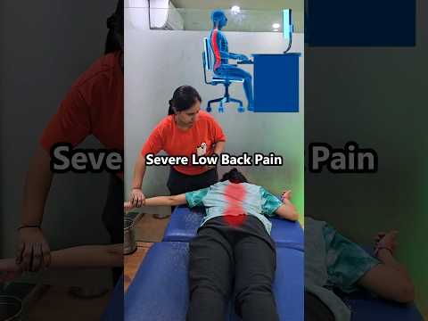
😭 Servere Low Back Pain due to Sitting #physiotherapy #chiropractor #lowerback
😭 Servere Low Back Pain due to Sitting #physiotherapy #chiropractor #lowerback
Frequently Asked Questions
Find clear answers about slip disc symptoms, diagnosis, treatment, and recovery.
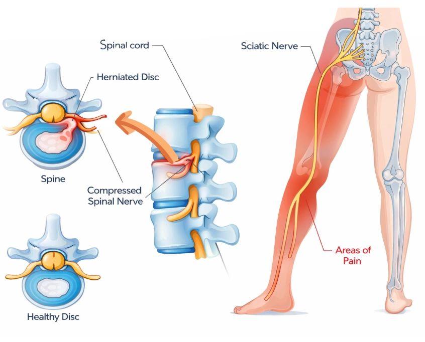
How disc herniation causes nerve pain
When the inner gel of a spinal disc pushes through its outer ring, it presses directly against sensitive nerve roots, creating sharp shooting pain...
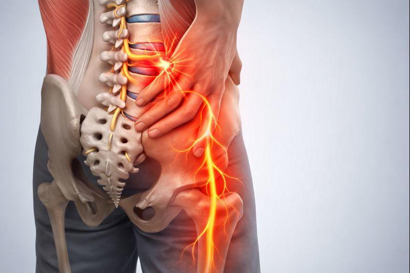
The connection to pelvic girdle pain
Lower lumbar nerve roots supply the pelvic and hip regions, meaning disc irritation can manifest as deep pelvic discomfort...
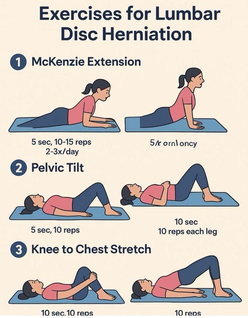
Natural healing and rehabilitation
With targeted physical therapy and proper movement guidance, most herniated discs retract and heal without requiring surgery...
When to See a Spine Specialist
Seek prompt care if you notice:
- Pain that doesn't improve after a few weeks of home care
- Worsening leg weakness or spreading numbness
- Loss of bladder or bowel control
- Pain severe enough to disrupt sleep or daily life
An early, accurate diagnosis makes slip disc treatment far more effective — relieving nerve pressure and restoring normal movement without unnecessary delay.
Book Spine Specialist Consultation