Hand Rehab: Tendon Cuts, Palsy , Wrist Drop & Stiffness
A detailed clinical guide to restoring strength, joint mobility, tendon gliding, and fine motor function following tendon cut injuries, radial/ulnar palsy, wrist drop, and post-fracture joint stiffness.
Book Free Consultation
Enter your details and our senior medical coordinator will reach out.
Comprehensive Hand & Nerve Rehabilitation
The hand's intricate network of tendons, nerves, and small joints requires specialized, structured rehabilitation following trauma or surgery. Conditions such as Tendon Cut Injuries, Radial Palsy, Ulnar Palsy, Wrist Drop, and Post-Fracture Stiffness demand precise, stage-appropriate therapy to prevent permanent contractures and muscle wasting.
Our clinic offers personalized hand rehab programs designed around your specific condition and surgical history, focusing on controlled tendon gliding, dynamic outrigger splinting, and progressive functional retraining.
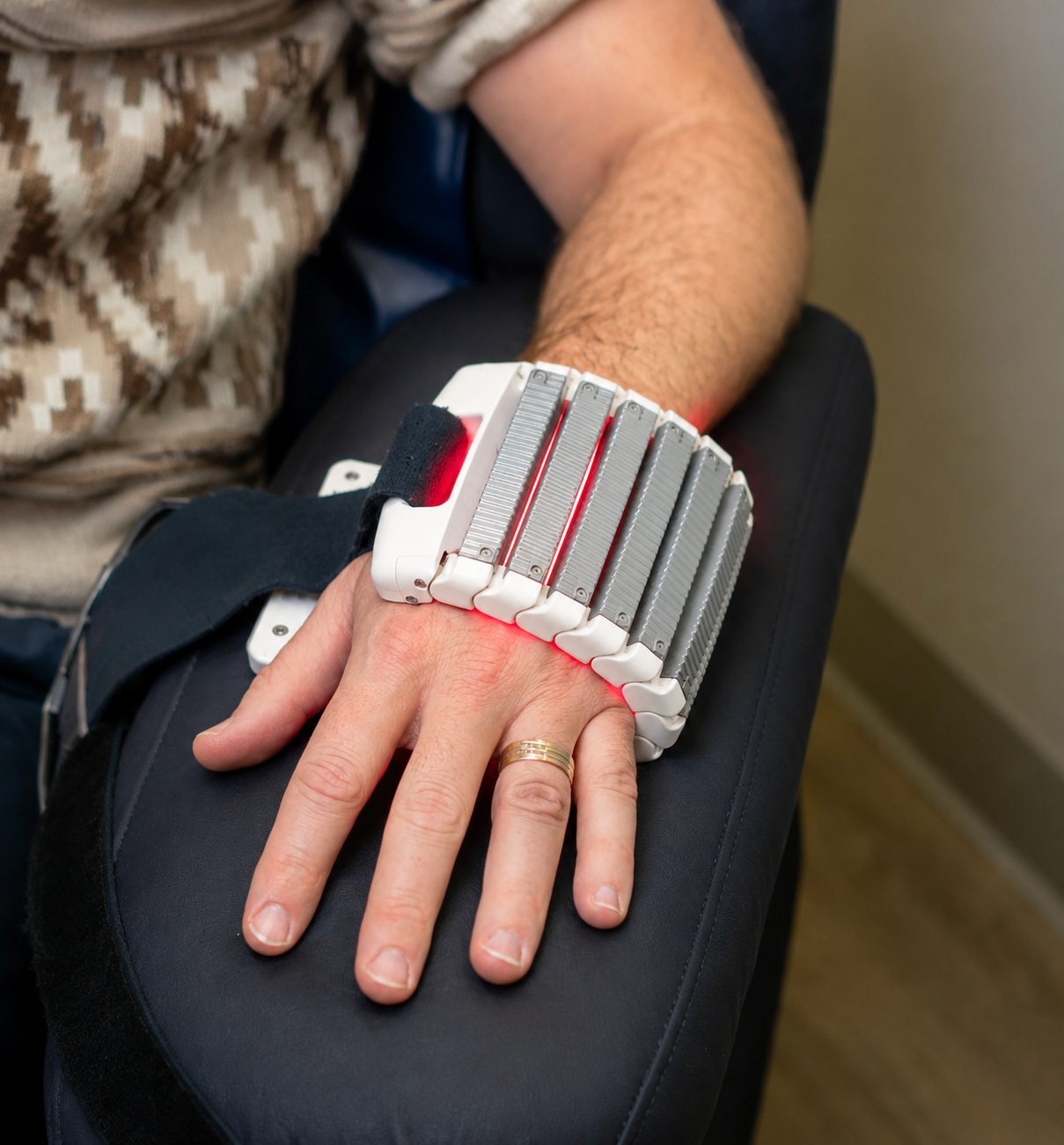
Symptoms of Hand Impairments & Nerve Deficits
Do not ignore these physical functional deficits following hand trauma, nerve palsy, or post-fracture immobilization:
Inability to Extend Wrist
Complete drop of the wrist and fingers due to radial nerve paralysis or wrist drop.
Claw Hand Deformity
Hyperextension at MCP joints and flexion at PIP/DIP joints from ulnar nerve palsy.
Tendon Laceration Lag
Inability to actively bend or straighten a specific finger following a tendon cut injury.
Post-Fracture Stiffness
Severe joint capsule tightness preventing full fist closure after cast removal.
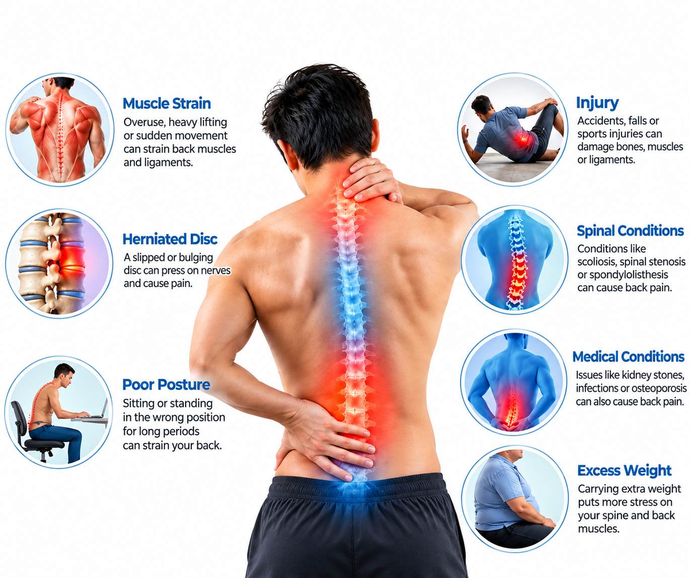
Causes of Hand Trauma, Palsy & Stiffness
Hand functional loss stems from traumatic lacerations, bone fractures, and compressive nerve injuries:
- Tendon Cut Injuries: Sharp glass or knife lacerations severing flexor or extensor tendons.
- Radial Palsy & Wrist Drop: Humeral shaft fractures, prolonged arm compression ("Saturday night palsy"), or nerve entrapment.
- Ulnar Palsy: Elbow trauma, cubital tunnel entrapment, or wrist lacerations affecting intrinsic muscles.
- Post-Fracture Stiffness: Prolonged immobilization of distal radius or metacarpal fractures leading to adhesion formation.
Treatment Strategy & Management Options
Managing these complex conditions ranges from dynamic outrigger splinting and early active motion to targeted strengthening:
Splinting & Tendon Gliding
Custom dynamic splints for wrist drop/palsy, controlled early active motion (CEAM) for tendon cut repairs, and scar mobilization.
Joint Mobilization & Strengthening
Serial casting and joint mobilization for post-fracture stiffness, combined with progressive grip strengthening and sensory re-education.
How Hand Trauma, Palsy & Stiffness Are Diagnosed
Our structured 4-tier clinical diagnostic protocol evaluates over 50 specific clinical and physical evaluation parameters for precise nerve palsy, tendon laceration, and stiffness assessment.
1. History, Surgical & Trauma Audit
Complete breakdown of tendon repair reports, fracture immobilization timelines, edema, and functional goals.
- Surgical Operative Report Review: Analyzing tendon repair zones (Zones I-V), nerve graft integrity, and plate fixation stability.
- Immobilization Timeline Audit: Tracking duration of post-fracture cast wear or nerve palsy onset.
- Edema & Swelling Assessment: Quantifying hand and finger fluid retention using volumetric measurements.
- Wound Healing & Incisions: Evaluating scar maturation and suture removal dates.
- Mechanism of Injury: Documenting glass laceration, crush trauma, humerus fracture, or Saturday night palsy.
- Occupational Hand Demands: Mapping fine motor and grip requirements for work return.
- Medication Efficacy Review: Documenting response to pain management during therapy.
- Visual Analog Scale (VAS): Recording resting and mobilization pain scores (0-10 scale).
- Baseline DASH Score: Measuring upper limb functional disability.
- Previous Therapy History: Reviewing prior exercises and patient compliance.
- Hand Dominance: Noting whether injured hand is dominant or non-dominant.
- Patient Recovery Goals: Aligning rehab milestones with personal or occupational pursuits.
- Surgeon Protocol Compliance: Adhering strictly to surgeon's motion restrictions.
- Psychosocial Barrier Screening: Evaluating fear of movement (kinesiophobia) post-injury.
2. Nerve Palsy vs Tendon Cut vs Stiffness Differential
Differentiating radial/ulnar nerve palsies from tendon lacerations and post-fracture joint contractures.
- Radial Nerve Palsy (Wrist Drop): Identifying inability to extend wrist/fingers while passive range remains full.
- Ulnar Nerve Palsy (Claw Hand): Differentiating intrinsic muscle paralysis (interossei/lumbricals) from tendon cuts.
- Tendon Cut Injury: Differentiating active lag from nerve paralysis using tenodesis action testing.
- Post-Fracture Joint Stiffness: Differentiating capsular contractures from muscle tendon tethering.
- Complex Regional Pain Syndrome (CRPS): Screening vasomotor changes and hyperalgesia post-fracture.
- Complete Sensory Mapping: Testing monofilament touch across radial, ulnar, and median distributions.
- EMG/NCS Correlation: Reviewing nerve regeneration electrodiagnostic reports for radial/ulnar palsies.
- Volkmann's Ischemic Contracture: Ruling out acute compartment syndrome sequelae post-fracture.
- Infection Screening: Ruling out osteomyelitis or deep wound infection post-surgery.
- Reflex & Strength Correlation: Matching clinical findings with nerve map.
- Joint Fusion vs Stiffness: Confirming whether restricted movement is articular or soft tissue.
- Tendon Rupture vs Adhesion: Assessing sudden loss of motion during rehab.
- Clinical Synthesis: Integrating all physical and history data.
3. Physical Exam & Special Tests
Total active motion (TAM), tenodesis action test, Froment's sign, grip dynamometry, and goniometry.
- Tenodesis Action Test: Passive wrist extension causing natural finger flexion (evaluating tendon/nerve gliding).
- Total Active Motion (TAM) Measurement: Summing active flexion minus extension deficits across digits.
- Froment’s Sign (Ulnar Palsy): Checking paper pinch between thumb and index for adductor pollicis weakness.
- Jamar Grip Strength Dynamometry: Testing power grip recovery post-fracture or repair.
- Pinch Gauge Testing: Evaluating tip-to-tip, key, and three-jaw chuck pinch strength.
- Scar Mobility Assessment: Palpating scar tethering and adhesion to underlying tendons.
- Active & Passive ROM Goniometry: Measuring MCP, PIP, and DIP joint angles to grade stiffness.
- Two-Point Discrimination Testing: Evaluating sensory acuity on fingertips post-nerve injury.
- Intrinsic Tightness Testing (Bunnell-Littler): Differentiating intrinsic muscle contracture from joint stiffness.
- Wartenberg's Sign: Checking little finger abduction in ulnar palsy.
- Nine-Hole Peg Test: Quantifying finger dexterity and coordination speed.
- Physical Baseline Record: Documenting TAM angles and grip kilograms for recovery tracking.
4. Splinting, Ultrasound & Care Roadmap
Custom thermoplastic splinting, dynamic outriggers, tendon gliding protocols, and functional rehabilitation.
- Custom Thermoplastic Splint Fabrication: Molding dynamic wrist drop outriggers or static extension splints for stiffness.
- Controlled Early Active Motion (CEAM): Implementing Kleinert/Duran protocols following tendon cut repairs.
- Scar Tissue Management & Desensitization: Silicone gel sheeting, elastomer pads, and deep scar massage.
- Edema Control Techniques: Retrograde massage and compression wrapping post-fracture.
- Sensory Re-Education Protocol: Textural training for reinnervating digital nerves post-palsy.
- Progressive Strengthening & Putty Training: Therapeutic putty exercises for muscle rebuilding.
- Multi-Specialist Hand Surgery Panel: Collaborative orthopedic hand surgeon and certified hand therapist review.
- Digital Archiving: Secure cloud storage of ROM charts and DASH progress scores in patient portal.
- Final Clinical Clearance: Approving independent return to work, cooking, and heavy manual activities.
- Dynamic Splint Adjustment: Modifying tension lines as joint mobility improves.
- Work Simulation Training: Practicing job-specific tool handling.
- Home Exercise Program Design: Tailoring independent stretching routines.
- Long-Term Joint Protection Briefing: Educating on joint conservation techniques.
Tendon Cut Injuries (Lacerations)
A tendon cut injury (laceration) occurs when sharp trauma (such as glass, knives, or machinery) severs flexor or extensor tendons in the hand, wrist, or forearm. Because severed tendons retract due to muscle pull, urgent surgical repair is required to reconnect the ends.
Following repair, the primary challenge in hand therapy is preventing dense scar adhesions from tethering the tendon to surrounding sheath tissues, which blocks finger movement. Our specialized protocols utilize Controlled Early Active Motion (CEAM) and relative motion splinting to safely promote tendon gliding during the critical healing window.
- Flexor & Extensor Lacerations: Managing zone-specific tendon repairs in the hand and fingers.
- Controlled Early Active Motion (CEAM): Balancing safe tension with immediate tendon gliding to eliminate scar adhesions.
- Protective Relative Motion Splinting: Keeping repaired fingers in a safe zone during initial weeks of healing.
Tendon Cut Injuries Key Focus Areas
- Surgical repair zone classification (Zones I-V)
- Kleinert or Duran controlled passive/active motion protocols
- Silicone gel sheeting and deep scar mobilization
- Progressive resistive strengthening following 8 weeks post-op
Radial Palsy
Radial Palsy occurs when the radial nerve is damaged, compressed, or paralyzed, resulting in the classic presentation of "wrist drop"—the complete inability to actively extend the wrist, fingers, or thumb. Common causes include humeral shaft fractures, prolonged compression from deep sleep ("Saturday night palsy"), or direct trauma.
Because peripheral nerves regenerate very slowly (roughly 1 millimeter per day), management focuses on protecting overstretched extensor muscles and maintaining passive joint flexibility using custom dynamic outrigger splints while awaiting nerve reinnervation.
- Nerve Paralysis & Wrist Drop: Managing complete loss of active wrist and finger extension.
- Dynamic Outrigger Splinting: Providing rubber-band assisted extension so patients can use their hand functionally during recovery.
- Nerve Regeneration Timeline: Monitoring slow recovery progress over 3 to 12 months.
Radial Palsy Key Focus Areas
- Custom thermoplastic dynamic wrist drop outrigger fabrication
- Preventing wrist and finger joint contractures through passive ROM
- Electrodiagnostic EMG/NCS tracking of nerve regeneration
- Progressive weaning from splints as active muscle control returns
Ulnar Palsy
Ulnar Palsy involves injury or entrapment of the ulnar nerve (often at the elbow or wrist), leading to paralysis of the intrinsic muscles of the hand (interossei and lumbricals). This causes a characteristic "claw hand" deformity where the ring and little fingers hyperextend at the knuckles and curl inward at the middle joints.
Patients lose critical lateral pinch strength (evaluated via Froment's sign) and dexterity. Our rehab protocols utilize anti-claw splints, intrinsic muscle strengthening, and sensory re-education to restore hand coordination.
- Intrinsic Muscle Paralysis: Managing interossei and lumbrical weakness in the hand.
- Claw Hand Deformity Correction: Utilizing anti-claw orthoses to block MCP hyperextension.
- Lateral Pinch Restoration: Rebuilding adductor pollicis strength and thumb stability.
Ulnar Palsy Key Focus Areas
- Froment's and Wartenberg's sign clinical examinations
- Anti-claw thermoplastic hand splinting
- Intrinsic strengthening and fine motor coordination drills
- Sensory re-education and protective hand education
Wrist Drop
Wrist Drop is a clinical sign rather than a standalone diagnosis, representing the inability to lift the wrist and fingers due to dysfunction of the radial nerve. When the wrist hangs limply in flexion, patients lose the ability to grasp objects effectively because optimal grip strength requires the wrist to be slightly extended.
Treatment requires immediate mechanical support via static and dynamic wrist extension splints to prevent overstretching of the paralyzed extensor muscles, combined with passive range of motion exercises and electrical stimulation to preserve muscle tone.
- Extensor Muscle Protection: Preventing irreversible overstretching of paralyzed forearm muscles.
- Functional Grasp Restoration: Utilizing wrist splints to enable stable grip mechanics.
- Electrical Muscle Stimulation: Maintaining muscle viability during prolonged nerve regeneration.
Wrist Drop Key Focus Areas
- Static cock-up and dynamic outrigger splinting
- Neuromuscular electrical stimulation (NMES) for forearm extensors
- Passive stretching to maintain full wrist and finger flexibility
- Graduated active strengthening upon nerve recovery
Post-Fracture Hand Stiffness
Post-Fracture Hand Stiffness is a frequent and disabling complication following wrist fractures (such as distal radius fractures) or metacarpal/phalangeal fractures requiring prolonged cast immobilization or surgical plate fixation.
When the hand is immobilized, joint capsules shrink, ligaments shorten, and soft tissues adhere, preventing full fist closure and finger straightening. Our specialized hand therapy protocols combine serial static-progressive splinting, paraffin wax heat therapy, joint mobilization, and active stretching to break down contractures and restore total active motion (TAM).
- Capsular Contracture Remodeling: Stretching shortened joint ligaments and capsules post-immobilization.
- Total Active Motion (TAM) Tracking: Measuring precise functional recovery of finger flexion and extension.
- Serial Progressive Splinting: Utilizing custom turnbuckle or progressive static splints to gain joint ROM.
Post-Fracture Stiffness Key Focus Areas
- Total Active Motion (TAM) and goniometric joint measurements
- Serial static-progressive and dynamic extension/flexion splinting
- Paraffin wax baths and manual joint mobilization techniques
- Progressive grip strengthening using therapeutic putty
Meet Our Hand Rehabilitation Experts
Specialized physiotherapy care focused on comprehensive Hand Rehabilitation, improving hand and wrist mobility, restoring grip strength and coordination, reducing pain and stiffness, and supporting a confident return to work, sports, and everyday activities through personalized rehabilitation.
Ask Our Specialist
Personalized hand rehabilitation focused on restoring hand and wrist mobility, improving grip strength and coordination, reducing pain and stiffness, enhancing functional control, and supporting a gradual return to comfortable everyday hand activities according to individual needs.
Treatment Videos/testimonials
Watch real recovery stories from patients who regained their active lifestyles.
No patient stories available.
Frequently Asked Questions
Clear answers to common clinical questions about tendon cut injuries, radial/ulnar palsy, wrist drop, and post-fracture stiffness.