Lumbar Spondylolisthesis: Causes, Symptoms and Treatment
A comprehensive medical guide to understanding vertebral slippage, spinal stability, diagnosis, and non-surgical care options.
Book Free Consultation
Enter your details and our senior medical coordinator will reach out.
What Is Lumbar Spondylolisthesis?
Lumbar spondylolisthesis occurs when a vertebra in the lower spine slips forward over the bone beneath it, out of its normal alignment. This shift can narrow the spinal canal and put pressure on nearby nerves, leading to lower back pain that often spreads into the legs.
It's graded by severity, from a minor slip to a more significant displacement requiring closer medical attention.
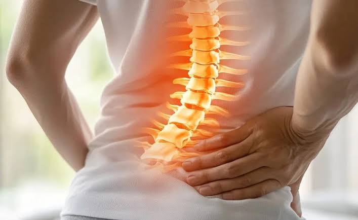
Common Symptoms
Do not ignore these physical indicators associated with vertebral slippage:
Persistent Back Pain
Lower back pain, often worse with standing or walking.
Forward Relief
Pain that eases when sitting or bending forward.
Tight Hamstrings
Tight hamstrings and reduced spinal flexibility.
Radiating Discomfort
Pain, tingling, or numbness radiating into the buttocks or legs.
What Causes It?
- A stress fracture in a vertebra (spondylolysis) — common in young athletes
- Age-related degeneration — of spinal discs and joints
- Repetitive strain — from sports involving bending or twisting
- Genetic factors — affecting bone strength and spine structure
- Traumatic injury — to the spine
- Excess body weight — adding pressure on the lower spine
How Lumbar Spondylolisthesis Is Diagnosed
Our structured 4-tier comprehensive diagnostic protocol maps over 80 specific clinical history, physical testing, and radiographic slip-grading parameters.
1. Medical History & Onset
Detailed recording of standing triggers, hamstring tightness, and slip history.
- Standing Pain Trigger: Evaluating lower back discomfort that worsens with prolonged standing.
- Walking Distance Tolerance: Recording claudication distance before lower back or leg pain forces rest.
- Forward Flexion Relief: Checking if sitting or bending forward relieves pressure.
- Hamstring Tightness Log: Recording chronic posterior thigh pull and restricted knee extension.
- Pain Radiation Patterns: Tracking shooting pain or numbness traveling into buttocks or calves.
- Onset Context: Determining if symptoms started during athletic hyperextension or gradual wear.
- Age & Athletic History: Evaluating youth sports involvement (gymnastics, football) or adult degeneration.
- Previous Spinal Trauma: Documenting historical falls onto buttocks or direct lower back impacts.
- Postural Change Reports: Noting self-observed changes in lordosis or pelvic tilt.
- Leg Weakness History: Recording episodes of giving-way or foot drag.
- Previous Treatments: Reviewing response to prior bracing, therapy, or medications.
- Family Spine History: Screening for hereditary spondylolysis or bone defects.
- Activity Limitations: Assessing how slippage impacts work, walking, and exercise.
- Weight & Load History: Checking mechanical body weight stress factors.
- Night Pain Review: Determining if instability disturbs sleeping posture.
- Bowel/Bladder Screening: Checking safety parameters for nerve root compression.
- Pain Intensity Scoring: Rating day-to-day stability pain using standard scales.
- Progression Rate: Evaluating how quickly standing tolerance has declined over months.
- Symptom Triggers: Identifying specific movements aggravating vertebral slip.
- Overall Health Overview: Comprehensive medical status summary.
2. Physical Examination
Hands-on instability testing, step-off palpation, and neurological screening.
- Step-Off Palpation: Checking for a palpable forward vertebral step deformity along spinous processes.
- Stork Test (Single-Leg Hyperextension): Screening for active pars interarticularis stress fracture.
- Hamstring Tightness Testing: Evaluating severe posterior chain restriction during straight leg raising.
- Posture & Alignment Check: Observing hyperlordosis and anterior pelvic tilt.
- Lumbar Range of Motion: Testing flexion, extension, and pain provocation angles.
- Neurological Screening: Testing deep tendon reflexes (patellar and Achilles).
- Myotome Muscle Testing: Assessing power in ankle dorsiflexion and big toe extension.
- Dermatome Sensory Exam: Screening for nerve root compression across lower limbs.
- Gait & Balance Assessment: Observing compensatory waddling or shortened stride.
- Segmental Instability Test: Applying gentle pressure to verify abnormal vertebral shifting.
- Core Muscle Strength: Evaluating abdominal bracing and lumbar support capacity.
- Hip Joint Screening: Ruling out hip impingement or referred groin pain.
- SI Joint Provocation: Testing for secondary pelvic joint strain.
- Trendelenburg Test: Checking hip abductor stability during single-leg stance.
- Toe and Heel Walking: Testing L5 and S1 motor root integrity.
- Myofascial Tension Check: Palpating paraspinal spasm secondary to instability.
- Flexibility Screening: Checking overall lower extremity muscle length.
- Stability Balance Test: Assessing trunk control under load.
- Functional Movement: Observing transition from sit-to-stand.
- Physical Summary: Compiling examination metrics.
3. Imaging & Slip Grading
Flexion-extension X-rays, MRI scans, Meyerding grading, and diagnosis.
- Standing Lumbar X-Rays: Essential upright views to capture vertebral displacement under gravity.
- Flexion-Extension X-Rays: Dynamic bending views to measure abnormal spinal instability.
- Meyerding Classification: Grading vertebral slip severity from Grade I (1-25%) to Grade IV (75-100%).
- MRI Scan Evaluation: Visualizing neural foraminal narrowing, disc health, and nerve root pinching.
- CT Scan & 3D Reconstruction: Detailed bony architecture view of pars defect (spondylolysis).
- Bone Scan (SPECT): Detecting active stress fracture healing in the pars interarticularis.
- Canal Stenosis Screening: Assessing neural canal dimensions during slippage.
- Blood Inflammatory Panels: Running ESR and CRP to rule out infectious or inflammatory spondylitis.
- Differential Diagnosis: Distinguishing degenerative slippage from isthmic or congenital causes.
- Nerve Conduction Studies: Assessing peripheral nerve damage from chronic root compression.
- Sequential Slip Tracking: Comparing past and present X-rays to check progression rate.
- Surgical Stability Review: Consulting spine surgeons for high-grade slips or progressive deficits.
- Osteoporosis Screening: DEXA scan for elderly patients presenting with degenerative slip.
- Digital Record Archiving: Secure cloud storage of radiological reports.
- Specialist Peer Review: Multi-expert consensus on slip management.
- Risk Stratification: Assessing nerve root tension risks.
- Prognosis Review: Estimating stability timelines.
- Lab Correlation: Matching systemic blood markers.
- Diagnostic Confirmation: Final report formulation.
- Comprehensive Clinical Diagnosis: Synthesizing physical instability tests, history, and dynamic X-rays into a final treatment roadmap.
4. Treatment & Recovery Plan
Targeted stabilization, core strengthening, and biomechanical care.
- Core Stabilization Regimen: Strengthening deep abdominal and lumbar muscles to brace the slipped vertebra.
- Flexion-Based Exercises: Prioritizing posterior pelvic tilt routines to reduce lumbar lordosis pressure.
- Hamstring & Hip Stretching: Safely relieving posterior chain pull on the pelvis.
- Postural Retraining: Correcting excessive anterior pelvic tilt during standing and walking.
- Bracing Support: Utilizing lumbar support corsets during acute instability flare-ups.
- Pain Management Care: Non-invasive modalities to alleviate muscular guarding and nerve irritation.
- Activity Modification: Advising avoidance of hyperextension sports and heavy axial loading.
- Home Exercise Program: Structured daily routines designed to maintain spinal control.
- Weight Management Support: Reducing mechanical shear stress on the lower lumbar joints.
- Ergonomic Workspace Setup: Optimizing chair and desk height to protect the lower back.
- Milestone Progress Tracking: Weekly clinical evaluations monitoring stability gains.
- Regular Clinical Follow-ups: Ongoing monitoring with senior spine care coordinators.
- Patient Self-Care Briefing: Clear instruction on managing slip flare-ups at home.
- Long-Term Protection Strategy: Preventing further vertebral translation through core endurance.
- Maintenance Care Routines: Sustaining spinal stability post-rehabilitation.
- Re-injury Prevention: Safe lifting mechanics training.
- Mobility Enhancement: Safe thoracic and hip movement preservation.
- Specialist Consult Coordination: Multi-expert review for advanced stability cases.
- Recovery Milestone Review: Assessing treatment success.
- Active Lifestyle Clearance: Final approval for unrestricted daily activities.
Treatment Options
Most mild to moderate cases of lumbar spondylolisthesis respond well to non-surgical treatment, with core strength and posture control playing a key role in long-term stability:
- Physiotherapy — to strengthen the core and stabilise the spine
- Activity modification — to avoid movements that worsen symptoms
- Posture correction and lower back support — when needed
- Pain-relief or anti-inflammatory medication — as advised by a doctor
- Epidural or targeted injections — for persistent nerve pain
- Surgery — considered only for severe slippage, significant instability, or unresolved nerve symptoms
Meet Our Spine Experts
Our lumbar spondylolisthesis care focuses on understanding vertebral slippage, managing lower back pain and leg discomfort, and supporting spinal stability. Our approach includes individual clinical assessment and a personalised rehabilitation plan designed to improve mobility and everyday function.
Ask Our Specialist
Care for lumbar spondylolisthesis may include clinical assessment, guided physiotherapy, core stabilization exercises, posture guidance, and activity modifications tailored to individual symptoms and everyday needs. Treatment recommendations are based on clinical evaluation and personal recovery goals.
Patient Success Stories
Explore patient videos and their details.
Patient Video
Select a video to view its details.
Patient Video
Select a video to view its details.
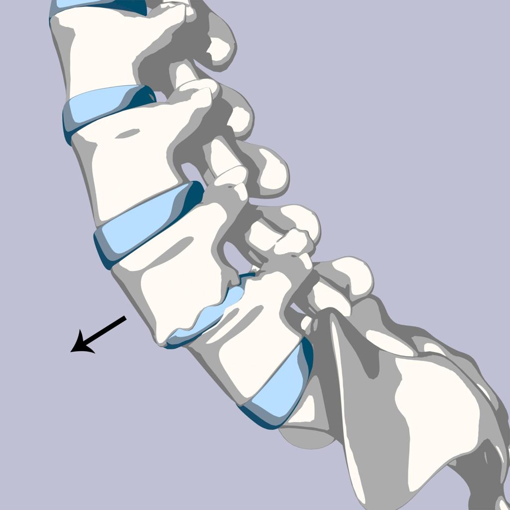
How does vertebral slippage cause leg pain?
When a vertebra slips forward, it narrows the spinal canal, compressing nerves that travel down into the legs...
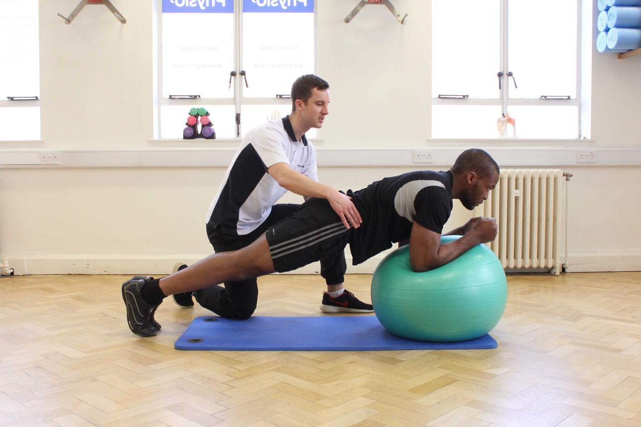
Why core strength stabilizes the spine
Targeted abdominal and back muscle conditioning provides a muscular brace, reducing movement at the slip site...
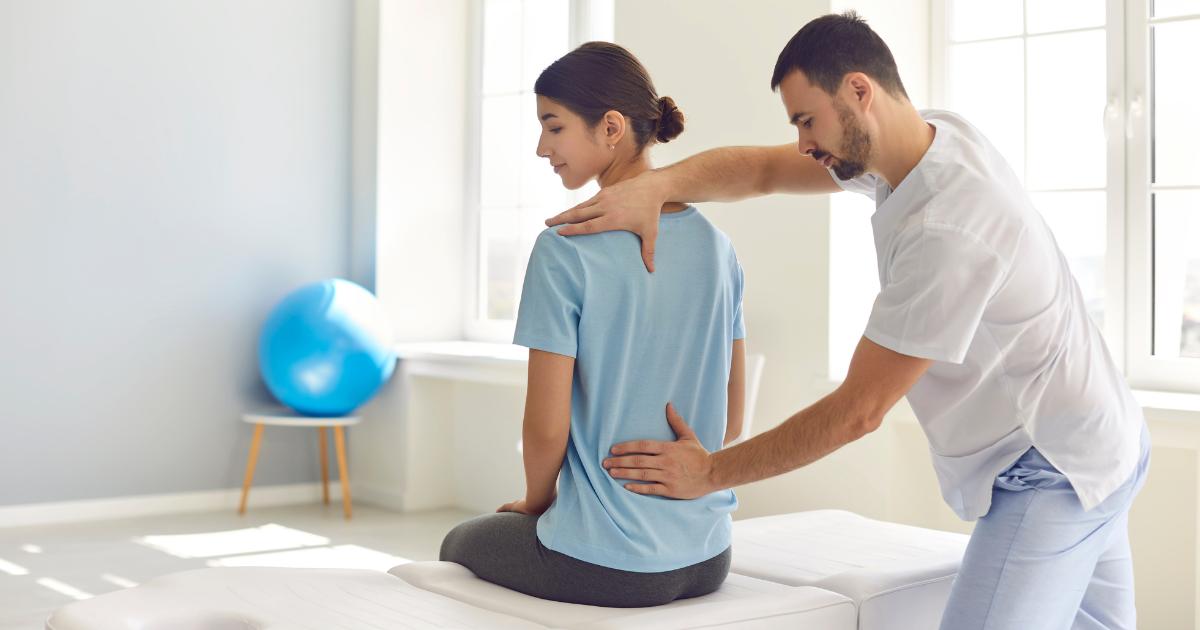
Understanding spondylolisthesis grades
Doctors grade the condition from Grade I to V based on the percentage of vertebral displacement over the bone below...
When to See a Spine Specialist
Seek medical evaluation for any of the following indicators:
- Back pain that persists or worsens over a few weeks
- Numbness, tingling, or weakness spreading into the legs
- Difficulty standing, walking, or maintaining balance
- Loss of bladder or bowel control
An early diagnosis helps prevent further vertebral slippage and keeps the spine stable, supporting faster recovery and better long-term mobility.
Book Spine Specialist ConsultationFrequently Asked Questions
Clear answers to common questions about lumbar spondylolisthesis, its symptoms, diagnosis, and treatment.