Foot & Heel Pain: Plantar Fasciitis, Calcaneal Spurs & Metatarsalgia
A comprehensive clinical guide to understanding plantar fascia inflammation, heel bone spurs, forefoot metatarsal overloading, precise diagnostic evaluations, and targeted non-surgical or surgical care options.
Book Free Consultation
Enter your details and our senior medical coordinator will reach out.
Understanding Plantar Fasciitis, Calcaneal Spurs & Metatarsalgia
Foot and heel pain can severely restrict your daily mobility, making every step a painful challenge. Three of the most common lower foot conditions include Plantar Fasciitis (inflammation of the thick band of tissue supporting the arch), Calcaneal Spurs (bony calcium growths on the heel bone often accompanying chronic fasciitis), and Metatarsalgia (pain and inflammation across the ball of the foot).
While plantar fasciitis causes sharp morning heel pain, metatarsalgia produces burning or aching across the forefoot heads. Our clinic provides specialized biomechanical evaluations, custom orthotic prescription, and targeted non-surgical care to restore normal weight distribution and relieve pressure across your entire foot.
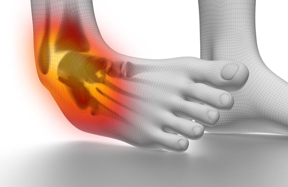
Symptoms of Plantar Fasciitis, Spurs & Metatarsalgia
Do not ignore these physical signs associated with arch strain, heel bone spurs, and forefoot overloading:
First-Step Morning Heel Pain
Sharp, stabbing pain in the heel during the very first steps out of bed or after sitting (Plantar Fasciitis).
Forefoot Ball Pain
Aching, burning, or sharp pain under the ball of the foot, often feeling like walking on pebbles (Metatarsalgia).
Chronic Heel Spur Ache
A persistent deep ache at the bottom center of the heel bone linked to traction bone spurs.
Activity Worsening
Pain that intensifies toward the end of the day or after prolonged standing, walking, or running.
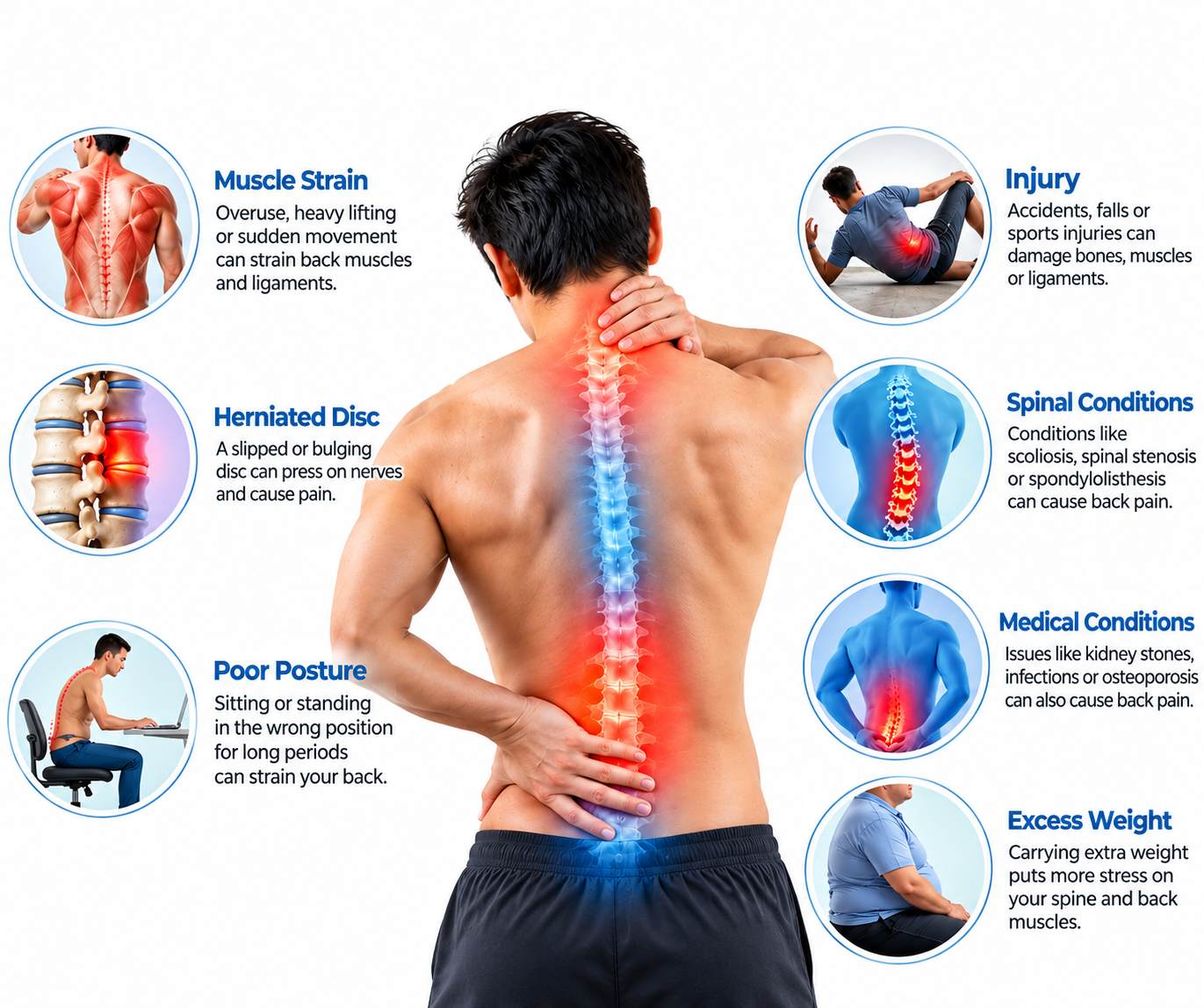
What Causes Plantar Fasciitis, Spurs & Metatarsalgia?
Arch inflammation, bone traction spurs, and forefoot pain stem from biomechanical overload, footwear, and high impact:
- Biomechanical Overpronation: Flat feet (pes planus) or high arches (pes cavus) placing excessive stretching tension on the plantar fascia.
- Chronic Traction Spurring (Calcaneal Spur): Long-standing plantar fascia pull on the calcaneus bone stimulating calcium deposition and spur formation.
- Forefoot Overloading (Metatarsalgia): High-heeled shoes, thin soles, or tight calf muscles shifting excessive body weight onto the metatarsal heads.
- Occupational Standing & Impact: Prolonged standing on hard concrete floors or sudden increases in high-impact running mileage.
Treatment Strategy & Foot Rehabilitation
Managing foot and heel pain centers on custom orthotics, night splints, shockwave therapy, and targeted stretching:
Orthotics, Night Splints & Stretching
Custom arch-support foot orthotics, dorsiflexion night splints, plantar fascia specific stretching, and footwear optimization.
Shockwave Therapy & Injections
Extracorporeal shockwave therapy (ESWT) for chronic heel spurs/fasciitis, metatarsal pads, and ultrasound-guided anti-inflammatory injections.
How Plantar Fasciitis, Spurs & Metatarsalgia Are Diagnosed
Our structured 4-tier clinical diagnostic protocol evaluates over 50 specific clinical and physical evaluation parameters for precise arch, heel, and forefoot diagnosis.
1. History, Footwear & Pain Pattern Audit
Complete breakdown of first-step morning heel pain, footwear choices, standing hours, and forefoot burning.
- First-Step Morning Heel Pain: Documenting classic sharp stabbing pain on first steps out of bed.
- Forefoot Burning / Pebble Feeling: Inquiring about aching ball-of-foot pain when walking barefoot (Metatarsalgia).
- Occupational Hard Flooring Hours: Measuring daily hours standing on concrete or tile.
- Footwear & High Heel Audit: Auditing use of unsupportive flat shoes, flip-flops, or high heels.
- Running Mileage & Surface Changes: Tracking recent increases in high-impact road running.
- Medication Efficacy Review: Documenting response to anti-inflammatory drugs or night splints.
- Visual Analog Scale (VAS): Recording morning, afternoon, and evening pain scores (0-10 scale).
- Baseline Foot Function Index (FFI): Measuring foot-related disability.
- Prior Foot Injury History: Screening past calcaneal bruises or stress fractures.
- Symptom Duration: Acute arch strain vs chronic calcaneal bone spurs.
- Previous Orthotic Trial: Assessing past use of arch supports or heel cups.
- Body Weight & BMI Audit: Evaluating weight load factors on plantar fascia.
- Patient Functional Goals: Aligning recovery with walking comfort and sports return.
- Bilateral Involvement Check: Assessing if one or both feet are symptomatic.
2. Fasciitis vs Calcaneal Spur vs Neuroma Differential
Differentiating plantar fasciitis and calcaneal spurs from metatarsalgia, Morton's neuroma, and calcaneal stress fractures.
- Plantar Fasciitis vs Calcaneal Spur: Calcaneal spurs are secondary bony projections at fascia insertion; plantar fasciitis is the actual soft tissue inflammation causing pain (spurs themselves rarely hurt directly).
- Metatarsalgia vs Morton's Neuroma: Metatarsalgia causes broad forefoot aching; Morton's neuroma causes sharp shooting nerve pain and numbness between 3rd/4th toes.
- Calcaneal Stress Fracture Rule-Out: Differentiating morning arch pain from deep calcaneal bone bruising or stress fractures.
- Baxter's Nerve Entrapment: Screening first branch of lateral plantar nerve entrapment mimicking plantar fasciitis.
- Fat Pad Atrophy Syndrome: Differentiating heel pad thinning in elderly from plantar fasciitis.
- Complete Neurological & Vascular Examination: Confirming normal tibial and sural nerve sensation.
- Inflammatory & Crystal Arthritis Screen: Ordering HLA-B27 and uric acid tests for ankylosing spondylitis or gout.
- Tarsal Tunnel Syndrome Screen: Ruling out tibial nerve compression behind medial ankle.
- Achilles Tendinitis Differentiation: Differentiating heel cord pain from plantar arch fascia pain.
- Infection / Osteomyelitis Rule-Out: Screening calcaneal bone infection.
- Referred Lumbar Radiculopathy (S1): Ensuring heel pain isn't radiating from lower back.
- Clinical Synthesis: Integrating physical tests with imaging.
3. Physical Exam & Special Tests
Windlass test, medial calcaneal tubercle palpation, metatarsal squeeze test, and ankle dorsiflexion ROM.
- Windlass Test (Plantar Fascia Gold Standard): Passively dorsiflexing big toe while foot is weight-bearing; reproducing sharp pain at medial calcaneal tubercle confirms plantar fasciitis.
- Medial Calcaneal Tubercle Palpation: Direct digital pressure pinpointing exact insertion tenderness of plantar fascia.
- Metatarsal Squeeze (Mulder's) Test: Compressing forefoot metatarsals together to rule out Morton's neuroma when evaluating forefoot pain.
- Weight-Bearing Ankle Dorsiflexion Lunge Test: Measuring calf/soleus flexibility limiting ankle motion.
- Foot Posture Index (FPI): Assessing flat feet (pes planus) vs high arches (pes cavus).
- Active & Passive ROM Goniometry: Measuring ankle and toe joint flexibility.
- Calcaneal Squeeze Test: Compressing heel bone laterally to screen calcaneal stress fractures.
- Forefoot Callus Inspection: Inspecting plantar skin pressure patterns under metatarsal heads.
- Gait & Stance Phase Observation: Evaluating arch flattening and push-off mechanics.
- Tinels Test at Tarsal Tunnel: Tapping tibial nerve behind medial ankle.
- Physical Baseline Record: Documenting Windlass test pain threshold and ankle dorsiflexion degrees for tracking.
4. Radiographs, Ultrasound & Care Roadmap
Foot and heel X-rays (calcaneal spur visualization), musculoskeletal ultrasound, shockwave therapy, and custom orthotics roadmap.
- Weight-Bearing Foot & Heel Radiographs (Lateral & AP): Visualizing calcaneal bone spurs (heel spurs), stress fractures, and metatarsal head alignment.
- High-Resolution Musculoskeletal Ultrasound: Dynamic imaging measuring plantar fascia thickness (>4mm indicates chronic fasciitis), fascial tearing, and surrounding inflammation.
- Extracorporeal Shockwave Therapy (ESWT): High-energy acoustic wave therapy stimulating cellular repair and neovascularization in chronic plantar fasciitis and heel spurs.
- Custom Foot Orthotics Prescription: Fabricating biomechanical arch supports to offload plantar fascia and redistribute pressure across metatarsal heads.
- Ultrasound-Guided Corticosteroid Injections: Precision anti-inflammatory infiltration into plantar fascia or metatarsal bursa for refractory pain.
- Night Splinting Protocol: Prescribing dorsiflexion night splints to maintain continuous passive stretch on plantar fascia during sleep.
- Multi-Specialist Podiatry & Orthopedic Panel: Collaborative podiatrist, orthopedic specialist, and physical therapist review.
- Digital Archiving: Secure cloud storage of foot X-rays, ultrasound logs, and FFI scores in patient portal.
- Final Clinical Clearance: Approving pain-free return to daily walking, running, and athletic footwear.
- Metatarsal Pad & Cushioning Guidance: Recommending forefoot offloading pads for metatarsalgia.
- Home Exercise Program Design: Tailoring independent arch stretching and calf lengthening routines.
- Long-Term Foot Health Briefing: Educating on supportive footwear and avoiding barefoot walking on hard surfaces.
Plantar Fasciitis
Plantar Fasciitis is the single most common cause of heel pain, involving inflammation of the plantar fascia—a thick, web-like ligamentous band connecting your heel bone to your toes that supports your foot arch. Patients classicly experience sharp, stabbing pain during the very first steps in the morning or after periods of sitting.
Driven by foot overpronation, tight calves, and prolonged standing on hard surfaces, our clinical protocols combine Windlass physical testing and musculoskeletal ultrasound with custom orthotics, night splints, and shockwave therapy to promote rapid healing.
- First-Step Morning Pain: Targeting the classic post-rest fascial tightening and micro-tearing.
- Windlass Mechanism Evaluation: Utilizing toe dorsiflexion to reproduce fascial tension.
- Night Splinting & Orthotics: Maintaining passive arch stretch and biomechanical support.
Plantar Fasciitis Key Focus Areas
- Windlass physical examination test for fascial tension
- Ultrasound measurement of plantar fascia thickness (>4mm)
- Custom orthotic arch support and dorsiflexion night splints
- Extracorporeal shockwave therapy (ESWT) for chronic pain
Calcaneal Spur (Heel Spur)
A Calcaneal Spur is a bony calcium projection (osteophyte) that develops on the underside of the heel bone (calcaneus) at the attachment site of the plantar fascia. Often visualized on lateral foot X-rays, heel spurs form in response to long-standing mechanical tension and chronic traction pull from the plantar fascia over many months or years.
Importantly, the bony spur itself is rarely the direct source of pain; rather, the surrounding soft-tissue inflammation of plantar fasciitis causes the discomfort. Management focuses on offloading fascial tension through custom orthotics, shockwave therapy, and targeted stretching rather than surgical spur removal.
- Traction Osteophyte Formation: Calcium deposition stimulated by chronic fascial pull on the calcaneus.
- X-Ray Visualization: Identifying hook-like bony projections on weight-bearing lateral radiographs.
- Soft-Tissue Focus: Treating underlying plantar fasciitis rather than targeting the bone spur directly.
Calcaneal Spur Key Focus Areas
- Weight-bearing lateral foot radiographs for spur identification
- Differentiating bony spur shadow from soft-tissue inflammation
- Shockwave therapy to stimulate healing at the insertion site
- Heel cushioning cups and custom orthotic pressure relief
Metatarsal Pain / Metatarsalgia
Metatarsalgia is a painful foot condition characterized by inflammation and aching across the ball of the foot (beneath the metatarsal heads). Frequently described as feeling like you are walking on pebbles or wearing a stone inside your shoe, it is common in runners, individuals wearing high heels, or those with altered foot mechanics.
Driven by excessive forefoot pressure loading, tight calves, or Morton's neuroma coexistence, management centers on metatarsal pad placement, footwear modification, cushioning orthotics, and calf stretching to redistribute body weight evenly across the foot.
- Forefoot Pressure Overload: Managing high compressive forces under the 2nd, 3rd, or 4th metatarsal heads.
- Mulder's Squeeze Testing: Differentiating mechanical metatarsalgia from Morton's interdigital neuroma.
- Metatarsal Pad Offloading: Redistributing forefoot weight using strategic insole padding.
Metatarsalgia Key Focus Areas
- Forefoot callus and pressure point clinical inspection
- Mulder's click test to rule out Morton's neuroma
- Metatarsal dome pads and custom cushioned orthotics
- Footwear modification (wide toe-box, low heel drop)
Meet Our Foot & Heel Specialists
Specialized care focused on managing Foot & Heel Pain, reducing discomfort during standing and walking, improving foot and ankle mobility, and supporting comfortable movement through personalized physiotherapy and rehabilitation.
Ask Our Specialist
Personalized Foot & Heel Pain rehabilitation focused on improving foot and ankle flexibility, strengthening supportive muscles, addressing walking mechanics, managing activity-related discomfort, and supporting a gradual return to comfortable everyday movement according to individual needs.
Treatment Videos/testimonials
Watch real recovery video from patients who overcame plantar fasciitis, heel spurs, and forefoot metatarsalgia.
No patient success stories are available right now.
Frequently Asked Questions
Clear answers to common clinical questions about plantar fasciitis, heel spurs, and metatarsalgia.