Young Age Knee Pain: PFPS, Chondromalacia & Patellar Alignment
A comprehensive clinical guide to anterior knee pain in teens and young adults, covering patellofemoral tracking disorders, retropatellar cartilage softening, Patella Alta and Patella Baja mechanics, and non-surgical restoration.
Book Free Consultation
Enter your details and our senior medical coordinator will reach out.
Understanding Knee Pain in Young Adults & Adolescents
Knee pain in adolescents and young adults is a distinct clinical challenge that should never be written off as mere "growing pains." The vast majority of cases stem from anterior patellofemoral imbalances, where the kneecap (patella) does not glide smoothly inside the femoral trochlear groove.
This spectrum encompasses Patellofemoral Pain Syndrome (PFPS), Chondromalacia Patella (softening and cracking of the retropatellar hyaline cartilage), and congenital or acquired patellar height anomalies such as Patella Alta (abnormally high-riding kneecap) and Patella Baja (abnormally low-riding kneecap). Our clinic provides thorough biomechanical evaluations and non-surgical restoration to stabilize patellar tracking.
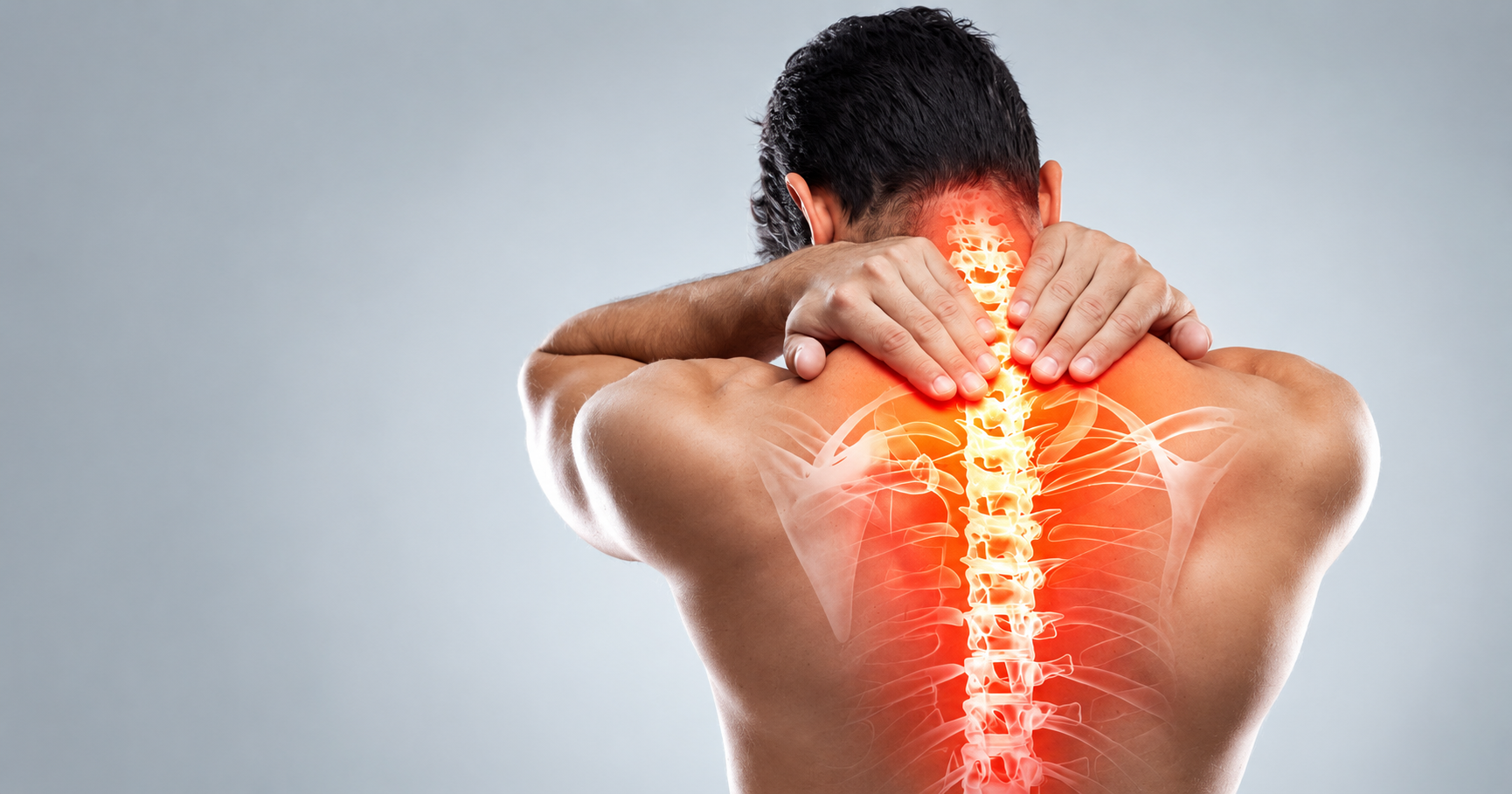
Symptoms of Young Age Knee Pain & Tracking Disorders
Do not ignore these physical signs indicating patellofemoral maltracking, cartilage irritation, or patellar height disorders:
Anterior Kneecap Ache
Diffuse aching behind or surrounding the patella, aggravated by squats, lunges, and stairs.
Moviegoer's Sign
Sharp stiffness and aching when standing up after prolonged sitting with knees flexed (theater sign).
Grating & Crepitus
Audible crunching, grinding, or cracking sounds during knee bending (Chondromalacia Patella).
Subluxation & Catching
A sensation that the kneecap is slipping out of place, giving way, or locking momentarily.
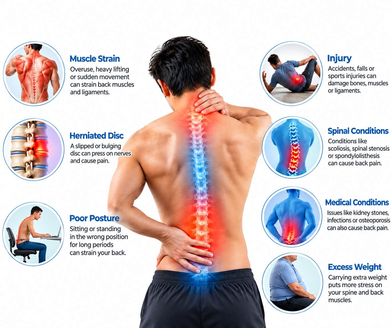
What Causes Young Age Knee Pain?
Anterior knee pain in youth stems from rapid skeletal growth, muscular imbalances, and structural patellar alignment anomalies:
- Quadriceps (VMO) & Hip Weakness: Weakness in the vastus medialis oblique and gluteus medius allowing the patella to track excessively lateral.
- Tight Lateral Retinaculum & IT Band: Excessive lateral tethering pulling the patella against the outer femoral trochlear ridge.
- Patella Alta (High Position): A long patellar tendon delaying engagement of the patella in the trochlea, causing recurrent instability.
- Patella Baja (Low Position): Shortened patellar tendon causing high compressive contact pressure and restricted knee flexion.
Treatment Strategy & Biomechanical Correction
Managing young knee pain centers on strengthening medial stabilizers, patellar realignment taping, and offloading cartilage:
VMO Activation & Hip Rehab
Closed-kinetic-chain quadriceps strengthening, gluteal neuromuscular retraining, and foam rolling of tight lateral structures.
McConnell Taping & Dynamic Bracing
Medial patellar glide taping to correct tracking, custom stabilizing knee sleeves, and orthotics to correct dynamic foot pronation.
How Young Age Knee Pain Is Diagnosed
Our structured 4-tier clinical diagnostic protocol evaluates over 50 specific clinical and physical evaluation parameters for precise patellofemoral tracking, cartilage condition, and height index assessment.
1. History, Growth & Patellofemoral Load Audit
Complete breakdown of growth spurt timing, sports participation, moviegoer's sign, and subluxation sensation.
- Adolescent Growth Spurt Correlation: Tracking sudden height changes creating quadriceps-hamstring muscle length discrepancy.
- Moviegoer’s / Theater Sign Audit: Noting stiffness and aching after sitting with knees bent at 90° during cinema or desk work.
- Stair Ascent & Descent Pain: Differentiating eccentric patellofemoral shear loading going downstairs.
- Patellar Subluxation Sensation: Documenting episodes where kneecap felt as if it slipped outwards or popped.
- Squatting & Kneeling Intolerance: Measuring threshold for deep knee flexion before anterior pain begins.
- Audible Crepitus & Grinding History: Inquiring if knee makes noisy grinding sounds during stair navigation.
- Sports & Dance Load Audit: Documenting participation in gymnastics, running, basketball, or ballet.
- Medication Efficacy Review: Recording response to anti-inflammatory pain medications.
- Visual Analog Scale (VAS): Recording resting, stair, and squatting pain scores (0-10 scale).
- Baseline Kujala Anterior Knee Pain Scale: Quantifying patellofemoral functional limitation score.
- Footwear & Arch History: Inquiring about flat feet (pes planus) collapsing knee inward.
- Symptom Duration: Acute traumatic dislocation vs chronic progressive tracking ache.
- Previous Taping or Bracing Trial: Assessing past response to patellar stabilizer braces.
- Patient Physical Activity Goals: Aligning recovery milestones with school physical education and sports return.
2. PFPS vs Chondromalacia vs Alta/Baja Differential
Differentiating functional maltracking from cartilage softening, structural height disorders, and meniscal lesions.
- PFPS vs Chondromalacia Patella: PFPS is functional maltracking without cartilage loss; Chondromalacia involves true structural softening and fissuring of retropatellar cartilage.
- Patella Alta vs Patella Baja: Alta features an elevated patella with late trochlear engagement and high dislocation risk; Baja features a low patella with severe compressive cartilage wear and restricted flexion.
- Osgood-Schlatter Disease Rule-Out: Differentiating patellofemoral pain from focal tibial tubercle growth plate apophysitis.
- Sinding-Larsen-Johansson Syndrome: Checking for traction apophysitis at the inferior patellar pole.
- Plica Syndrome Differentiation: Differentiating medial parapatellar synovial shelf entrapment from generalized PFPS.
- Meniscal Tear Exclusion: Ruling out true mechanical joint line locking and McMurray clicks.
- Osteochondritis Dissecans (OCD) Screen: Checking for loose bone-cartilage fragments in femoral condyles.
- Referred Hip Pain Check (SCFE/Perthes): Ensuring adolescent knee pain is not radiating from an undiagnosed hip condition.
- Complete Neurological & Vascular Examination: Confirming normal lower extremity motor power and sensation.
- Inflammatory Arthritis Screening: Ordering ESR and CRP to rule out juvenile idiopathic arthritis.
- Hoffa’s Fat Pad Impingement: Differentiating fat pad pinch from retropatellar cartilage irritation.
- Septic Arthritis Emergency Rule-Out: Ruling out hot, swollen infected joint emergencies.
- Clinical Synthesis: Integrating all physical and history data.
3. Physical Exam & Special Tests
Clarke's test, J-sign patellar tracking, Q-angle measurement, patellar tilt, and apprehension test.
- Clarke’s Test (Patellar Grind Test): Downward pressure on superior pole of patella while patient contracts quadriceps reproducing retropatellar pain.
- J-Sign Dynamic Tracking Test: Observing lateral deviation of kneecap as knee extends from 90° to full extension (indicates Patella Alta / lateral maltracking).
- Patellar Apprehension (Fairbank's) Test: Pushing patella laterally at 20-30° flexion testing for patient anxiety or guarding (instability/subluxation).
- Q-Angle Measurement (Goniometry): Measuring angle formed between ASIS, patellar center, and tibial tubercle (elevated >20° increases lateral pull).
- Patellar Tilt Test: Attempting to lift lateral edge of patella; inability to lift to horizontal confirms tight lateral retinaculum.
- Patellar Glide Assessment: Measuring medial/lateral translation in quadrants (excessive medial glide in Patella Alta, restricted glide in Patella Baja).
- Vastus Medialis Oblique (VMO) Atrophy Inspection: Inspecting medial muscle bulk and timing of contraction.
- Single-Leg Squat Assessment: Observing dynamic knee valgus collapse and pelvic drop during loading.
- Active & Passive ROM Goniometry: Measuring knee flexion limits (often severely restricted in Patella Baja).
- Ober’s Test: Evaluating IT band and TFL tightness tethering the lateral patella.
- Hamstring & Calf Flexibility Testing: Checking popliteal angle and ankle dorsiflexion.
- Physical Baseline Record: Documenting Q-angle, patellar quadrant glide, and Kujala functional scores for tracking.
4. Radiographic Indices, MRI & Care Roadmap
Insall-Salvati ratio, Caton-Deschamps index, Merchant skyline views, 3T MRI cartilage mapping, and targeted care roadmap.
- Lateral Knee Radiograph (Insall-Salvati Ratio): Measuring ratio of patellar tendon length to patellar diagonal length (>1.2 confirms Patella Alta; <0.8 confirms Patella Baja).
- Caton-Deschamps & Blackburne-Peel Indices: Secondary radiographic ratios verifying patellar height relative to tibial plateau.
- Merchant / Laurine Skyline Radiographs (30-45° Flexion): Visualizing congruence angle, sulcus angle, and lateral patellar tilt.
- High-Resolution 3T Knee MRI (T2 Cartilage Mapping): Detecting early retropatellar cartilage softening, swelling, and fissuring (Chondromalacia grades I-IV).
- Trochlear Dysplasia Assessment: Evaluating flat or shallow femoral groove anatomy on axial MRI.
- Tibial Tubercle-Trochlear Groove (TT-TG) Distance: Measuring lateral displacement vector (>20mm indicates high instability risk).
- Targeted McConnell Patellar Taping Protocol: Clinical taping to correct lateral glide, tilt, and rotation.
- Closed-Kinetic-Chain Physiotherapy Protocol: Pain-free leg presses, step-downs, and terminal knee extensions.
- Multi-Specialist Orthopedic & Sports Medicine Panel: Collaborative sports physician, knee surgeon, and physical therapist review.
- Digital Archiving: Secure cloud storage of Insall-Salvati X-rays and MRI cartilage maps in patient portal.
- Final Clinical Clearance: Approving pain-free return to high-impact running, competitive sports, and school activities.
- Custom Foot Orthotics Prescription: Correcting overpronation to normalize dynamic Q-angle.
- Surgical Realignment Triage: Surgical evaluation (MPFL reconstruction, lateral release, or tibial tubercle osteotomy) for recurrent dislocations.
Patellofemoral Pain Syndrome (PFPS)
Patellofemoral Pain Syndrome (PFPS), commonly called "runner's knee" or anterior knee pain syndrome, is the most common cause of knee pain in adolescents, runners, and young active adults. It occurs when abnormal tracking causes the patella to rub unevenly against the femoral trochlear groove during knee flexion and extension.
Unlike structural cartilage damage, PFPS is primarily a biomechanical disorder driven by dynamic knee valgus, delayed Vastus Medialis Oblique (VMO) firing, and weak hip abductors. Patients classicly present with anterior aching after prolonged sitting ("moviegoer's sign") and pain going downstairs.
- Dynamic Knee Valgus Correction: Strengthening gluteus medius to eliminate inward knee collapse during running.
- VMO Neuromuscular Retraining: Restoring medial pull on the kneecap to prevent lateral tracking.
- McConnell Realignment Taping: Providing immediate mechanical unloading of irritated patellar facets.
PFPS Key Focus Areas
- Eliminating moviegoer's sitting knee stiffness
- VMO activation and closed-chain quadriceps loading
- Dynamic single-leg squat alignment screening
- Foot orthotics to correct excessive subtalar pronation
Chondromalacia Patella Syndrome
Chondromalacia Patella involves true structural deterioration, softening, and fissuring of the thick hyaline cartilage lining the posterior underside of the kneecap. Often developing as a progression of long-standing, untreated PFPS maltracking, it represents true retropatellar chondropathy.
Graded from Stage I (cartilage softening) to Stage IV (deep erosion exposing subchondral bone), patients experience pronounced retropatellar crepitus, grinding, and sharp pain during deep squats. Our clinic utilizes advanced 3T MRI cartilage mapping and viscosupplementation or regenerative therapies to protect and preserve articular integrity.
- Outerbridge Staging: Grading chondromalacia from mild softening (Grade I) to bone exposure (Grade IV).
- Retropatellar Cartilage Protection: Avoiding open-chain deep knee extensions that spike patellofemoral compressive stress.
- Viscosupplementation & PRP: Enhancing joint lubrication and stimulating native chondrocyte preservation.
Chondromalacia Key Focus Areas
- Clarke's patellar grind provocation testing
- 3T MRI T2 mapping to evaluate cartilage thickness and fissures
- Joint-friendly low-impact rehabilitation (cycling/swimming)
- Anti-inflammatory and lubricating joint therapy
Patellar Dysfunction: Patella Alta (High-Riding Kneecap)
Patella Alta is a structural condition where the kneecap sits abnormally high above the femoral trochlear groove, typically defined radiographically by an Insall-Salvati ratio greater than 1.2. This occurs due to an unusually long patellar tendon (patella ligament).
Because the patella sits so high, it does not enter the stabilizing bony trochlear groove until the knee is bent past 30 degrees of flexion. In early flexion, the kneecap floats loosely on the soft tissues, resulting in dynamic J-sign tracking, severe patellofemoral instability, and recurrent lateral patellar dislocations in adolescents.
- Delayed Trochlear Engagement: The kneecap remains unstable during the initial 0-30° of knee flexion.
- Recurrent Dislocation Risk: Extremely high incidence of lateral patellar subluxations in active teenagers.
- Stabilizing Rehabilitation: Intensive VMO strengthening and stabilizing knee bracing to prevent lateral dislocation.
Patella Alta Key Focus Areas
- Insall-Salvati ratio > 1.2 on lateral knee radiographs
- J-sign dynamic tracking observation upon full extension
- Patellar stabilization braces with lateral buttress supports
- Medial Patellofemoral Ligament (MPFL) integrity assessment
Patellar Dysfunction: Patella Baja (Low-Riding Kneecap)
Patella Baja (also known as Patella Infera) is the opposite anatomical anomaly, where the kneecap rests abnormally low on the femur, defined radiographically by an Insall-Salvati ratio of less than 0.8. It frequently develops secondary to patellar tendon contracture or scarring following knee trauma, surgery, or prolonged immobilization.
A low-riding kneecap causes continuous, extremely high compressive contact pressures across the patellofemoral joint even at rest. This leads to marked restriction of knee flexion range of motion, severe anterior knee pain, early-onset patellofemoral osteoarthritis, and quadriceps extensor mechanism disruption.
- Severe Joint Contact Pressure: Immense compressive loading between patella and lower trochlea.
- Marked Flexion Deficit: Inability to fully bend the knee due to patellar tendon shortening and tethering.
- Aggressive Mobilization Protocols: Intensive patellar mobilization, scar release, and extensor lengthening.
Patella Baja Key Focus Areas
- Insall-Salvati ratio < 0.8 on lateral knee radiographs
- Superior patellar glide mobilization to restore tendon length
- Aggressive post-surgical scar desensitization and release
- Preventing premature bone-on-bone patellofemoral arthritis
Meet Our Young Knee Care Experts
Specialized care focused on Young Age Knee Pain, identifying movement-related concerns, improving knee mobility and strength, addressing muscular imbalances, and supporting healthy functional movement through personalized physiotherapy and rehabilitation.
Ask Our Specialist
Personalized rehabilitation for young individuals experiencing knee pain, focusing on strengthening supporting muscles, improving flexibility and joint control, addressing posture and movement patterns, and managing activity-related discomfort. Treatment goals support a gradual return to comfortable daily activities and sports participation.
Treatment Videos/testimonials
Watch real recovery videos from patients who regained their active lifestyles.
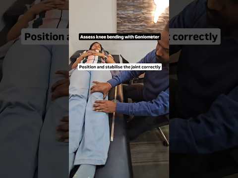
🤯 Goniometer का कमाल | How to use Goniometer to asses knee bending | #physiotherapy #chiropractor
Patient experience and recovery journey
🤯 Goniometer का कमाल | How to use Goniometer to asses knee bending | #physiotherapy #chiropractor
Patient experience and recovery journey
Frequently Asked Questions
Clear answers to common clinical questions about young age knee pain, PFPS, Chondromalacia, and Patella Alta/Baja.